Macular degeneration also been known as the age-related macular degeneration (AMD or ARMD) is an eye pathological condition that affect the macular area causing the cone photoreceptor cells within the macular area to be died off.
Retina is the interior layer of the eye which is extremely sensitive toward light. It consists of a few layers with bundle of nerve network connection and rich with blood supply as well. At the central portion of retina, lies the macula area which responsible for detail fine vision as well as diferrentiate color vision. At these area, the photoreceptors that densely packed are known as the cone photoreceptor cells. Surrounding the retina will be the peripheral fundus which lies the rod photoreceptor cells. Rods mainly use to sense the motion and moving thing around and helps us to see better in a dimmed light condition.
In ARMD patient, their central vision will be affect with a black spot (commonly known as the scotoma) but the peripheral vision remain intact. Hence, it is normal for a ARMD patient to tilt their head a bit to see a person face because at a normal straight position, their view will be obstructed making them unable to distinguish a person face. The main reason that cause ARMD will be owing to the increased age whereby the macula cells are deterioted (innevitable physiological process). Although this condition may cause significant vision problems, it never leads to complete blindness as it affects only the central part of the vision and the side usually, or peripheral vision is always left undisturbed.

In term of differentiates ARMD, it can be divided into two types. The first one will be the dry ARMD which is the degeneration of the cells in the retina with deposition of drusen at macula. Generally, most of the ARMD patient will begin in the dry form (estimated approxiamtely 80% of ARMD patient suffer from dry type ARMD). This type of ARMD results from the gradual wither of the retinal cells at the macular. As the cell are deterioted, drusen will be seen to be deposited on these area making its one of the sign to be diagnosed as dry ARDM. These spots are located in the back of the eye at the level of the outer retina and are able to be seen using an ophthalmoscope or a slit-lamp biomicroscope. Advanced dry macular degeneration, known as geographic atrophy, is the culmination of prolonged, progressive wasting changes in the nerves and sensory retina which main the cause of of vision loss in dry ARMD, not drusen.
For the second type, it is the wet ARMD which is caused the abnormal ingrowth of new blood vessels underneath the retina layer (choroidal neovascularisation, CNV). These newly growth blood vessel are very weak and fragile and easily leak causing bleeding to be occur inside the macular area. This type of haemorrhages will subsequetly cause distortion and damage to the centre of retina. Compared to the dry type of ARMD, wet ARMD will cause a faster vision loss and normally one eye will be affected first before rapidly affect the other eyes. Althought the wet ARMD are found in 20% of among ARMD patient, it takes into accounts for almost two-thirds of the people who have significant visual loss.
Sign and Symptom
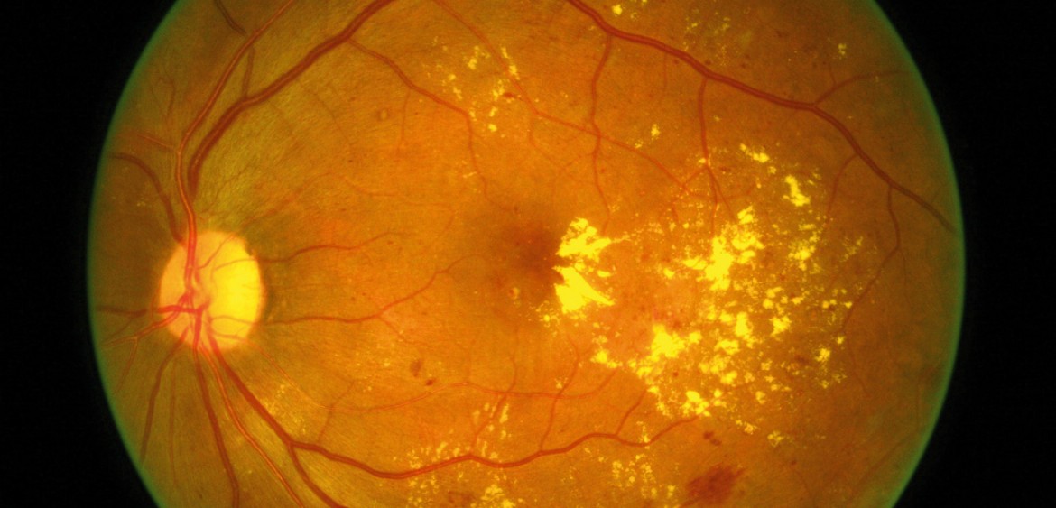
For the sign of ARMD patient, they may exhibit reduced vision (distance and near) and most of them can be categorized as low vision patient if they having impairment of visual functioning even after treatment, and/ or standard refractive correction, and has a visual acuity of less than 6/18 to light perception or a visual field of less than 10 degrees from the point of fixation, but who uses, or is potentially able to use, vision for the planning and/or execution of a task. By viewing through their fundus, ARMD patient’s fundus will show a degenerated macular area with darker area color at the macular side compared to the surrounding fundus area.
Patient with ARMD will normally complaint of blurry central vision to a total loss of central vision. Due to the degeneration of the macular area (mostly the cone photoreceptor area) they will also having a reduction or total loss of color perception and contrast sensitivity as well. Along with this, ARMD patient will suffer from photophobia but they will see better at night or in dimly lit areas due to usage of rod cells system ( surrounding the fundus area) to perceive. Thus, ARMD patient will required a longer recovery period when the light illumination level are changing from bright illuminated toward dimly illuminated. The depth of perception maybe affected as well. For some typical cases, such as the patient with wet type of ARMD, they may having a distortion of vision which are the things that they perceived may appear to be wavy or irregular (metamorphopsia). This features can be diagnosed using the Amsler Grid chart which mainly used to screen the macular central field of view.
Management
As an optometrist, patient with ARMD condition should be inform and explain to them about the vision problem they currently facing. A patient with deppressed problem or emotional psychological condition may not help in improving their vision at all. Hence, the patient must first accept their eye condition and have a positive attitude and motivation as well during the session course of rehabilitation.
The most common technique that teach to ARMD patient is the eccentric fixation. This is an alternative way of using the residual vision (peripheral vision) from the surrounding area besides from the damaged macular area which is still intact. For some patient, eccentric fixation is the best option to help them regain back some of the ability to see and focus at far and distance. But this require the technique and training in order to make it successful.
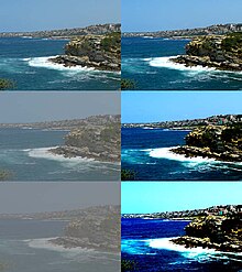
Besides that, optometrist can also prescribe tinted or absorptive lenses to help patient with outdoor activities. Some color of tints have been designed, and are marketed specifically, to improve the comfort and visual performance of visually impaired people who suffered from a range of ocular disorders such as ARMD, retinitis pigmentosa (RP), cataract, cone dystrophy and oculo-cutaneous albinism. It was found that short wavelength light has been shown to cause visual discomfort, hazy vision, reduced contrast and prolonged adaptation times. Hence, some of the tinted prescriptive lenses can actually helps to alleviate some or all of these effects by filtering out the blue light (short wavelength) in the visible portion of the spectrum that create problems for the photophobic or ageing eye. Yet, there are other type of prescriptive lenses with a other tinting such as yellow, amber or grey filter that can be helps to improve contrast and control glare or photophobia problem as well. For yellow and orange lenses, they helps a lot in increasing the contrast sensitivity of the ARMD patient. Grey lenses helps to reduce the loss of contrast sensitivity usually suffered in the presence of glare.
As usual, low vision aid devices will be last resort that prescribed to improve or enchance the residual or potential vision that might still left in ARMD low vision patient. There are different type of variety low vision aid device availabe in this market but not necessary its all work for all the ARMD low vision patients. Some ARMD low vision patient might find the low vision aid devices are helpful and useful depending on the situation they using the devices and the condition of their eye vision as well. Some of the example of low vision aid devices which can be prescribed to ARMD low vision patients are such as hand-held magnifier, spectacle incorporated addition magnifier, reversed telescope, amorphic lenses, negative lens, fresnel prism, mirror and field-expanding channel lens. All these low vision aid devices have its own mechanism to function and some are used to see the distance and some are used to see the near. Optometrist can prescribe these low vision aid devices depending on the habit and needs of the ARMD low vision patients. Most importantly, patient attitude and motivation will determine the rate of successful in using the low vision aid devices.
Finally, ARMD patient should always refer to their optometrist in order to monitor their eye condition continuously.
To have more information, please feel free to consult with our best optometrists in KL.